Frequently Asked Questions
- Who is eligible to host a deployment of the IHS-JVN Teleophthalmology Program?
- How do I apply for hosting a deployment of the IHS-JVN Teleophthalmology Program?
- What costs does the IHS-JVN hosting site bear?
- Who can be an imager?
- What is involved in the training for the imager?
- How much time is required for imaging duties each day?
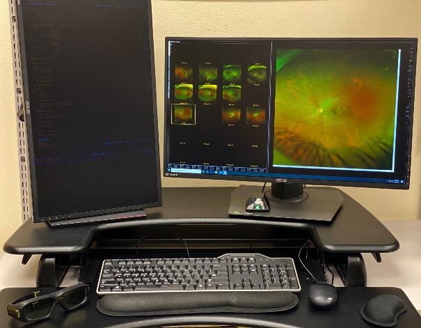
- Who owns and maintains the IHS-JVN equipment?
- Where do I get technical and clinical support for operating the program?
- What type of patient should undergo imaging?
- How are patients recruited for imaging?
- Who reads the images?
- Is credentialing and privileging required at the hosting site?
- Does the Centers for Medicare and Medicaid Services (CMS) or The Joint Commission (TJC) have any special requirements for supplying this telemedicine service at our facility?
- How are reports obtained from the Telemedicine Program?
- How can I know that the JVN system is safe and effective?
- Would a live examination be better than JVN imaging?
- Does and IHS-JVN examination replace a complete eye exam?
- Usually a diabetic eye examination requires dilation of the patient's pupil with eye drops. Does the JVN system use eye drops?
- Will the JVN system produce a change in our eye department workload?
- How is the IHS-JVN Teleophthalmology Program funded?
Q: Who is eligible to host a deployment of the IHS-JVN Teleophthalmology Program?
A: Any Indian Health Service, Tribal, or Urban Indian Health Program (I/T/U) is eligible for a deployment of the IHS-JVN. Specific sites selected for deployment are determined based upon site interest, capacity, public health criteria, and IHS policy.
Q: How do I apply for hosting a deployment of the IHS-JVN Teleophthalmology Program?
A: Send a message through the Contact Us section of our website requesting consideration for participation in the program and supply the following information:
- Facility name and location
- Point of Contact
- Name
- Title
- Phone Number
- Email Address
Q: What costs does the IHS-JVN hosting site bear?
A: The hosting site supplies the salary for the imager and travel costs for training/certification at the Phoenix Indian Medical Center. This is almost always a part-time position. The hosting site must also supply the space to house the Imaging Acquisition Station.
All other direct costs of the deployment and operation of an IHS-JVN Imaging Acquisition Station are borne by the IHS-JVN Teleophthalmology Program itself. This includes the following:
- All Equipment, software, and services directly related to imaging and transfer of the images
- Camera, imaging computer
- JVN/Chronic Disease Management Program (CDMP) software
- Technical and clinical support
- Maintenance/repairs
- Reading and reporting services
- Quality assurance (QA)
- Initial and recurrent Training and certification of Imager
Q: Who can be an imager?
A: A special background or previous training is not requisite for imager trainees. This skill level for this task is defined in a Position Description classified at GS6-7, and is well within the capacity of an existing clerk. Previous medical or nursing training is not required, but may be helpful. More important than the background of the imager is the immediate availability of the imager, since this is frequently a fractional FTE duty. The imager should be selected based upon interest and consistent availability to the imaging station.
Q: What is involved in the training for the imager?
A: All imagers are trained to demonstrated proficiency. This typically requires two full days and occurs at the IHS-JVN offices in Phoenix, AZ. Most of the imagers are Native Americans, and training at PIMC allows them to train in an IHS facility on IHS patients with a Native American trainer.
Q: How much time is required for imaging duties each day?
A: The total time required by the imager is generally dependent upon the size of the active diabetic registry and the ability of your staff to effectively recruit patients for IHS-JVN imaging. If imaging is available every ordinary work day, approximately 2 hours of imaging per day is needed for every 600 patients with diabetes. Additional administrative time is needed for scheduling and retrieving reports.
Q: Who owns the IHS-JVN equipment?
A: The IHS-JVN Teleophthalmology Program retains ownership of all equipment and software supplied by the program. The IHS-JVN Teleophthalmology Program maintains the equipment and updates the software as needed.
Q: Where do I get technical and clinical support for operating the program?
A: The IHS-JVN Teleophthalmology Program clinical and technical support via a help desk as needed.
Q: What type of patient should undergo imaging?
A: The primary purpose of the IHS-JVN Teleophthalmology Program is to increase the annual diabetic retinopathy examination rate. At a minimum, all patients with diabetes who have not had an eye examination within the past year should undergo IHS-JVN Imaging. Patients must carry a diagnosis of diabetes, either type one or type two to be eligible for imaging. Patients who are pregnant and had pre-existing diabetes prior to pregnancy are also eligible. Pre-diabetic patients are not eligible for imaging.
Q: How are patients recruited for imaging?
A: The initial goal of the IHS-JVN Teleophthalmology Program is to increase the annual diabetic retinopathy rate at its hosting site. The patients should be recruited from the clinic where your patients obtain primary care of their diabetes. The most efficient method targets all patients with diabetes. At a minimum all patients with diabetes who have not had an eye examination within the past year should be selected from your primary care or diabetes clinic for IHS-JVN imaging. This is best done using Electronic Health Record or iCare tools.
Q: Who reads the images?
A: Certified JVN Optometrist readers at the IHS-JVN National Reading Center in Phoenix interpret the retinal images.
Q: Is credentialing and privileging required at the hosting site?
A: Imager:
Your Imager will obtain training and provisional certification by a JVN imager trainer. Imaging services can be delivered by your imager staff based upon job description or Position Description, and doesn't require privileging except as required by your Medical Staff Bylaws.
Reader:
The JVN images are read and reported by a trained and certified Reader at the IHS-JVN National Reading Center. This reader is a licensed optometrist or ophthalmologist that is privileged for clinical and telemedicine services at the Phoenix Indian Medical Center (PIMC). The IHS-JVN readers also should be privileged at your facility via by Proxy credentialing. This is done using the Centers for Medicare and Medicaid Services (CMS) method of Reliance Privileging of telemedicine providers. This process relies upon the credentialing process of PIMC, but privileging of each IHS-JVN provider is still granted by your Governing body.
Q: Does the Centers for Medicare and Medicaid Services (CMS) or The Joint Commission (TJC) have any special requirements for supplying this telemedicine service at our facility?
A: CMS and THC standards require that telemedicine providers be credentialed in your facility. This is provided by CMS Reliance Privileging of telemedicine providers. While many TJC standards apply to telemedicine as a component of general healthcare service delivery, LD LD.04.03.09 and MS.13.01.01 apply specifically to telemedicine.
Q: How are reports obtained from the Telemedicine Program?
A: The JVN report is currently available to the hosting site through a web based report retrieval.
Q: How can I know that the JVN system is safe and effective?
A: The JVN has been tested in controlled studies that have passed the scrutiny of academic peer review. These studies have clearly demonstrated the validity of the JVN program as compared to the Early Treatment Diabetic Retinopathy Study (ETDRS). Since the ETDRS is the gold standard for evaluating diabetic retinopathy, the JVN is considered safe and effective. These studies include but are not limited to the following:
Bursell SE, Cavallerano JD, Cavallerano AA, Clermont AC, Birkmire-Peters D, Aiello LP, Aiello LM; Joslin Vision Network Research Team. Stereo nonmydriatic digital-video color retinal imaging compared with Early Treatment Diabetic Retinopathy Study seven standard field 35-mm stereo color photos for determining level of diabetic retinopathy. Ophthalmology 2001;108:572-85.
Silva PS, Cavallerano JD, Sun JK, Noble J, Aiello LM, Aiello LP. Nonmydriatic ultrawide field retinal imaging compared with dilated standard 7-field 35-mm photography and retinal specialist examination for evaluation of diabetic retinopathy. American Journal of Ophthalmology 2012;154:549-59.
Olvera-Barrios A, Heeren TF, Balaskas K, Chambers R, Bolter L, Tufail A, Egan C, Anderson J. Comparison of true-colour wide-field confocal scanner imaging with standard fundus photography for diabetic retinopathy screening. British Journal of Ophthalmology 2020 Mar 4. pii: bjophthalmol-2019-315269. doi: 10.1136/bjophthalmol-2019-315269. [Epub ahead of print].
Q: Would a live examination be better than JVN imaging?
A: The conventional and most prevalent method of evaluating diabetic patients for retinopathy has been a dilated retinal examination by an ophthalmologist or optometrist. However, the gold standard for establishing the level of diabetic retinopathy is a photographic method established by the Early Treatment Diabetic Retinopathy Study (ETDRS). IHS-JVN has been proven to have equivalency with the ETDRS. For this reason, an IHS-JVN examination may be viewed as equivalent to a conventional live examination for the purpose of detecting diabetic retinopathy.
Q: Does and IHS-JVN examination replace a complete eye exam?
A: No; a periodic complete eye examination is a component of good health care for almost everyone. The IHS-JVN examination is equal or better than a live dilated retinal examination for the purpose of achieving standard of care for diabetic retinopathy surveillance and diagnosing diabetic retinopathy, but it does not replace other features of a complete eye exam such as a check of intraocular pressure or glasses. However, it does meet/exceed the standard of care needed to avoid the consequence of the leading cause of new blindness among working age adults, diabetic retinopathy. A comprehensive eye examination with dilation may still be needed for e.g. pre-existing eye condition other than DR, a glaucoma evaluation, need for new glasses/contact lenses, periodic general eye evaluation, etc.
Q: Usually a diabetic eye examination requires dilation of the patient's pupil with eye drops. Does the JVN system use eye drops?
A: The IHS-JVN system uses a specially designed camera that does not require pupil dilation in most cases. In certain cases with unusually small pupils a very weak and short acting drop may be used to improve image quality.
Q: Will the JVN system produce a change in our eye department workload?
A: Yes. Most health care facilities in the IHS are able to provide diabetic retinopathy examinations to approximately 50% their known patients with diabetes each year. Participating in the IHS-JVN Teleophthalmology Program can substantially increase this examination rate. Referrals from the program must receive a live examination, so any effectively deployed IHS-JVN system would be expected to increase the patient load on the system used to provide eye examinations. However, when properly implemented, the program reduces the number of routine diabetic retinopathy exams as patients diagnosed with minimal or no diabetic retinopathy will not be referred to the eye clinic. The net change is one of increased efficiency rather than increased workload.
Q: How is the IHS-JVN Teleophthalmology Program funded?
A: The IHS-JVN Teleophthalmology Program is funded by appropriation language that provides funding specifically for this purpose. This language specifies the use of the IHS-JVN system.